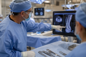
Live fluoroscopy with a C-arm is critically important in spine, pain management, and orthopedic procedures because it provides real-time X-ray imaging during surgery or needle placement, allowing clinicians to see anatomy, instruments, implants, and contrast flow as the procedure happens.
That real-time visualization greatly improves accuracy, safety, and procedural success.
What a C-arm fluoroscopy system does
A C-arm is a mobile imaging system shaped like the letter “C” that produces continuous or pulsed X-ray images during procedures.
It allows surgeons and interventional physicians to:
- visualize bones and joints dynamically,
- guide needles and implants precisely,
- confirm anatomical positioning,
- monitor movement during manipulation,
- and immediately detect complications.
Why it matters so much in spine procedures
The spine contains:
- the spinal cord,
- exiting nerve roots,
- major blood vessels,
- and very small anatomical target zones.
Even a few millimeters of error can cause:
- nerve injury,
- paralysis,
- bleeding,
- chronic pain,
- or failed surgery.
Examples where live fluoroscopy is essential
Pedicle screw placement
In spinal fusion surgery, screws must pass through narrow pedicles without breaching bone.
Live fluoroscopy helps surgeons:
- verify trajectory,
- monitor depth,
- avoid the spinal canal,
- and confirm hardware placement.
Without imaging guidance, malposition rates and neurological risks increase significantly.
Vertebroplasty and kyphoplasty
During cement injection into vertebrae, fluoroscopy allows clinicians to monitor cement flow in real time.
This is critical because cement leakage into:
- the spinal canal,
- veins,
- or lungs
can cause catastrophic complications.
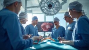
Disc procedures and minimally invasive spine surgery
Fluoroscopy guides:
- tubular retractors,
- dilators,
- endoscopes,
- and decompression tools
through very small incisions.
The surgeon often cannot directly see deep anatomy externally, so imaging becomes the “eyes” of the procedure.
Why fluoroscopy is vital in pain management
In interventional pain procedures, physicians place needles extremely close to:
- nerves,
- epidural spaces,
- joints,
- and blood vessels.
Blind injections are much less reliable and carry greater complication risk.
Epidural steroid injections
Live fluoroscopy helps confirm:
- exact spinal level,
- needle depth,
- and contrast spread.
Contrast dye can show whether medication is entering:
- the epidural space correctly,
- a blood vessel accidentally,
- or the wrong tissue plane.
This dramatically improves both safety and treatment accuracy.
Nerve blocks and radiofrequency ablation
Procedures targeting tiny medial branch nerves or sympathetic chains depend on highly accurate needle positioning.
Fluoroscopy improves:
- reproducibility,
- lesion precision,
- and procedural effectiveness.
Why it is essential in orthopedic surgery
Orthopedic procedures often require exact mechanical alignment.
Fracture fixation
During trauma surgery, fluoroscopy allows surgeons to:
- reduce fractures in real time,
- align bone fragments,
- place rods, screws, and plates,
- and verify fixation stability.
This reduces:
- malalignment,
- limb deformity,
- and repeat surgeries.
Joint procedures
In hip, pelvis, and extremity procedures, fluoroscopy helps with:
- implant positioning,
- leg-length assessment,
- hardware orientation,
- and joint congruity.
Intramedullary nailing
Passing a rod through the marrow canal requires continuous imaging to:
- avoid cortical perforation,
- maintain alignment,
- and place locking screws accurately.
Major safety advantages
Live fluoroscopy helps prevent:
- wrong-level spine surgery,
- vascular injection,
- nerve injury,
- misplaced hardware,
- organ injury,
- and failed procedures.
In many cases, it directly reduces medicolegal and catastrophic complication risk.
Why “live” imaging matters more than static X-rays
Static imaging only provides snapshots.
Live fluoroscopy allows clinicians to:
- see movement dynamically,
- adjust trajectory instantly,
- observe contrast flow in motion,
- and respond immediately to unexpected findings.
That continuous feedback is especially important when anatomy shifts during breathing, positioning, or surgical manipulation.
Relationship to newer robotic and navigation systems
Modern robotic spine and orthopedic systems often still rely heavily on fluoroscopy.
The imaging may be integrated with:
- computer navigation,
- 3D reconstruction,
- AI-assisted targeting,
- or robotic guidance platforms.
Fluoroscopy acts as a foundational imaging input for many advanced technologies.
The tradeoff: radiation exposure
One important limitation is radiation exposure to:
- patients,
- surgeons,
- staff,
- and trainees.
That is why modern practice emphasizes:
- pulsed fluoroscopy,
- low-dose protocols,
- collimation,
- shielding,
- distance techniques,
- and minimizing beam time.
Even with these concerns, the benefits of accurate image guidance usually far outweigh the risks in properly indicated procedures.
Bottom line
Live fluoroscopy with a C-arm is so important because it transforms many spine, pain, and orthopedic procedures from partially “blind” interventions into highly visualized, precision-guided operations.
It improves:
- anatomical accuracy,
- procedural confidence,
- patient safety,
- surgical outcomes,
- and complication prevention,

![]()